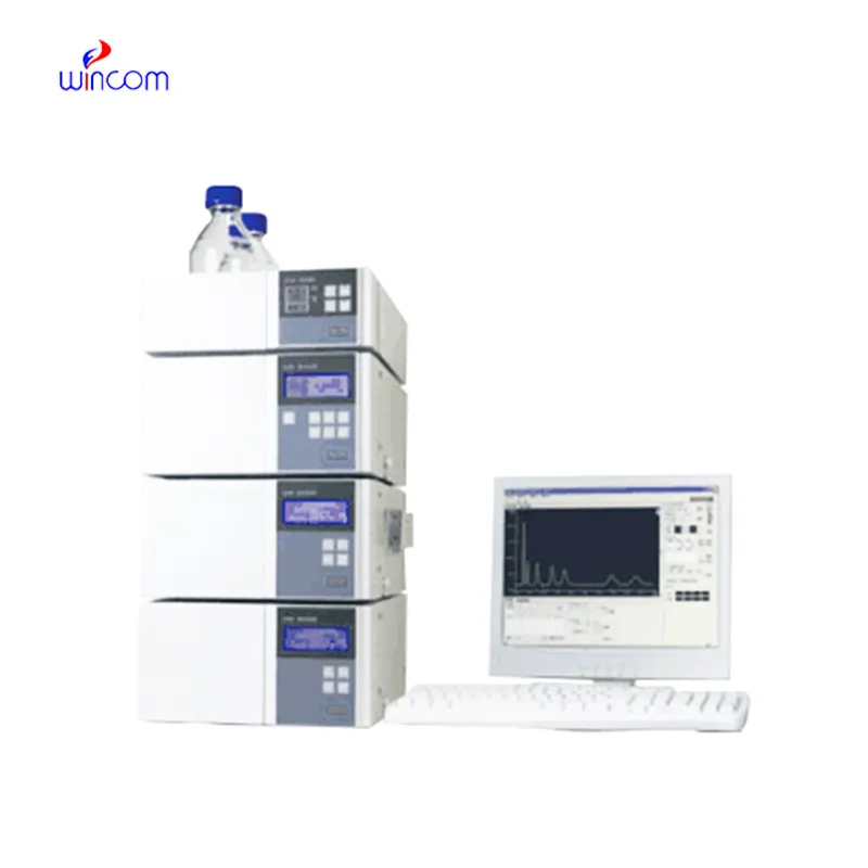
Amino Acid Liquid Chromatography is a critical technique to obtain analytical information in studies of medicines, clinical samples, and biochemistry. It isolates compounds according to their chemical characteristics, generating reproducible analytical results. Laboratory scientists use Amino Acid Liquid Chromatography to perform drug stability tests, monitor patient biomarkers, and find impurities. Its very high accuracy and flexibility allow thorough sample analysis in research, hospital, and clinical laboratory environments, thus becoming a fundamental device for assuring precision in both experimental and diagnostic results.

The hospital laboratory technicians employ Amino Acid Liquid Chromatography to quantify the quantity of proteins or peptides. This assists in the research of biomarkers, immunotherapy studies, and analysis of responses induced by novel therapies among patients. Its accuracy and sensitivity enable the obtaining of correct results, hence aiding superior research.
The forthcoming breed of Amino Acid Liquid Chromatography will put a spotlight on intelligent instruments that are connected with cloud-based surveillance. Through this monitoring, hospitals will be able to gain a remote view of laboratory activities and the results of sample analysis. Lab productivity will be greatly increased by the upcoming Amino Acid Liquid Chromatography, and together with the new features, patient testing and therapy monitoring even in difficult clinical settings will be more accurate.
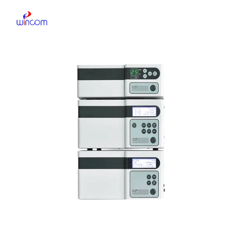
The effectiveness of a laboratory is determined by the proper maintenance of Amino Acid Liquid Chromatography. If the pump seals are regularly cleaned, the flow rates are monitored, and the usage of incompatible solvents is avoided then damage to the laboratory equipment can be prevented. It is essential for the technicians to carefully examine the columns, detectors, and tubing and in case of any sign of wear to conduct the scheduled calibration. Keeping Amino Acid Liquid Chromatography in their best condition guarantees reproducibility, lowers the risk of equipment breakdown, and provides continuous performance for both hospital tests and experiments.
Amino Acid Liquid Chromatography is equipped with an in-depth examination of biomolecules like proteins, peptides, and nucleic acids. Reversed-phase, ion-exchange, and size-exclusion chromatography methods qualify scientists to get insight into the molecular properties with utmost accuracy. The application of Amino Acid Liquid Chromatography in metabolomics studies, enzyme kinetics, and protein characterization helps in high accuracy and reproducibility. The high sensitivity level helps to detect low-molecular-weight molecules in detail and get insight into biological samples at a high level. One of the prime reasons why scientists are interested in Amino Acid Liquid Chromatography is its ability to generate information that advances understanding at an advanced biochemistry level.
Q: What is HPLC used for in laboratories? A: HPLC turns out to be one of the most significant and essential analytical methods in laboratories equipped with the chemical compound analysis, separation, identification, and quantification of their presence in complex samples which are the research, clinical, and pharmaceutical applications. Q: How does HPLC separate compounds? A: The HPLC separation technique is based on the different affinities of the compounds to the stationary phase and mobile phase within the chromatography column. Q: Can HPLC analyze biological samples? A: Yes, it is certainly possible to carry out analyses on various biological fluids such as blood, serum, urine, etc. for the detection of metabolites, drugs, and biomarkers. Q: How often should HPLC columns be replaced? A: The replacement of the columns must be done according to the manufacturer instructions or when the performance begins to decline, which is quite usual after heavy use or contamination. Q: What detectors can be used with HPLC? A: The analysis type determines the use of, among others, UV, fluorescence, refractive index, and mass spectrometry detectors as the common detectors.
The microscope delivers incredibly sharp images and precise focusing. It’s perfect for both professional lab work and educational use.
The hospital bed is well-designed and very practical. Patients find it comfortable, and nurses appreciate how simple it is to operate.
To protect the privacy of our buyers, only public service email domains like Gmail, Yahoo, and MSN will be displayed. Additionally, only a limited portion of the inquiry content will be shown.
We’re interested in your delivery bed for our maternity department. Please send detailed specifica...
We are planning to upgrade our imaging department and would like more information on your mri machin...
E-mail: [email protected]
Tel: +86-731-84176622
+86-731-84136655
Address: Rm.1507,Xinsancheng Plaza. No.58, Renmin Road(E),Changsha,Hunan,China

 af
af
 es
es
 ar
ar
 tr
tr
 sw
sw
 pt
pt
 th
th
 ur
ur
 bn
bn
 ne
ne
 vi
vi
 km
km
 lo
lo
 de
de
 ru
ru
 fi
fi
 nl
nl
 fa
fa
 fr
fr
 ko
ko