The branching hyphae under microscope combines technological innovation and practical design, with distortion-free, clear-quality imaging at every magnification. The mechanical stability and focus precision controls of the branching hyphae under microscope ensure accurate specimen positioning. The branching hyphae under microscope enhances sample visibility in varying light conditions using a strong illumination system. Optional camera adapters and measuring software are offered to extend its use, making it suitable for various scientific and educational environments.
The branching hyphae under microscope is critical to science and manufacturing advancement. In the medical research arena, the branching hyphae under microscope aids microscopic blood and tissue testing for accurate diagnostics. Research institutions use the branching hyphae under microscope in cell culture analysis, detecting bacterial growth, and research on biofilms. Industrial laboratory environments utilize the branching hyphae under microscope for product quality assurance and surface finishes evaluation. The branching hyphae under microscope is also applied in environmental science to support monitoring of plankton populations and particles of pollutants, to enhance ecological studies and sustainability science.

The future of the branching hyphae under microscope is influenced by digitalization and smart automation. More efficient imaging sensors will allow the branching hyphae under microscope to identify three-dimensional structures with unprecedented precision. Artificial intelligence will analyze microscopic images, reduce human errors, and optimize research productivity. Wireless communication and cloud connectivity will facilitate collaboration globally with remote monitoring and immediate data exchange. The branching hyphae under microscope will be an entirely networked instrument that closes the gap between laboratory precision and data-driven research outcomes.

The branching hyphae under microscope has the strength of longevity, which is dependent on the right handling and maintenance by cleaning regularly. Clean the eyepieces, objectives, and stage with accepted lens paper after each use. Remove all slides and samples prior to shutdown. The branching hyphae under microscope should be stored in a cool, dry place to avoid corrosion and mold. Check screws and mechanical joints for support at intervals. The electrical components, such as the power supply unit and light source, should be inspected frequently to ensure safe operation.
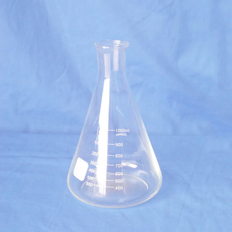
A branching hyphae under microscope is able to closely study microorganisms, tissue, and materials and is thus a fundamental instrument in laboratories and classrooms. It operates by bending light or electron rays to enlarge specimens to appear gigantic many times magnification. The branching hyphae under microscope has been enhanced with developments in optics to enable brighter, clearer, and digital-imaging-assisted magnification. In academic research work as well as industrial inspection, a branching hyphae under microscope enables accurate analysis, recording, and examination of complex microscopic realms.
Q: What is the lifespan of a microscope? A: With proper care and maintenance, a microscope can last for many years, providing consistent optical performance and stability. Q: How does the objective lens affect image quality in a microscope? A: The objective lens determines magnification and resolution; high-quality lenses produce sharper, more accurate images of specimens. Q: Can a microscope be used to view live specimens? A: Yes, many microscope models support live-cell observation, allowing users to study biological processes in real time under controlled conditions. Q: What is the function of the condenser in a microscope? A: The condenser focuses light onto the specimen, enhancing illumination and improving contrast for clear image viewing. Q: How should a microscope be transported safely? A: Carry the microscope with both hands—one under the base and one on the arm—to prevent damage or misalignment of delicate parts.
The delivery bed is well-designed and reliable. Our staff finds it simple to operate, and patients feel comfortable using it.
We’ve used this centrifuge for several months now, and it has performed consistently well. The speed control and balance are excellent.
To protect the privacy of our buyers, only public service email domains like Gmail, Yahoo, and MSN will be displayed. Additionally, only a limited portion of the inquiry content will be shown.
I’d like to inquire about your x-ray machine models. Could you provide the technical datasheet, wa...
We’re looking for a reliable centrifuge for clinical testing. Can you share the technical specific...
E-mail: [email protected]
Tel: +86-731-84176622
+86-731-84136655
Address: Rm.1507,Xinsancheng Plaza. No.58, Renmin Road(E),Changsha,Hunan,China

 af
af
 es
es
 ar
ar
 tr
tr
 sw
sw
 pt
pt
 th
th
 ur
ur
 bn
bn
 ne
ne
 vi
vi
 km
km
 lo
lo
 de
de
 ru
ru
 fi
fi
 nl
nl
 fa
fa
 fr
fr
 ko
ko