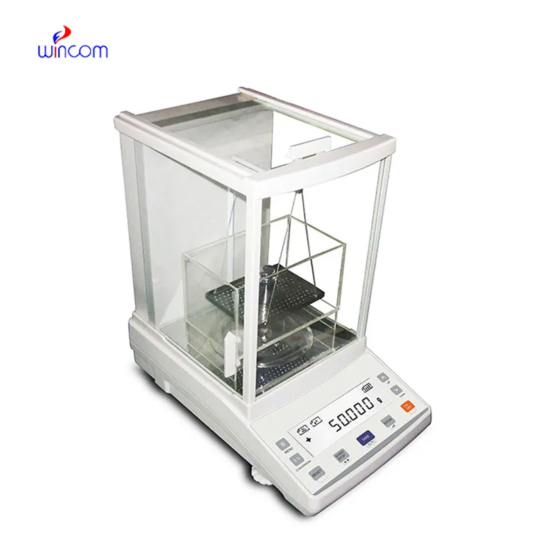
The use of digital electronic analytical balance in pharmaceutical laboratories is crucial for the accurate measurement of both active substances and excipients. Its extraordinary accuracy eliminates the possibility of formulation errors and makes regulatory compliance easier. digital electronic analytical balance are employed by laboratory staff for daily quality control, validation of batches, and research activities. Adding digital electronic analytical balance to the laboratory operations not only the consistency but also the reproducibility and the accuracy of the results for clinical trials and research applications are assured.

In research labs of the biomedical field, digital electronic analytical balance is used while standardizing the experimental samples. For the purpose of testing, h researchers have to measure the biological or chemical samples very accurately and in this way, they do not use more than the required amount of sample for analytical testing. This process keeps the studies that compare different methodologies consistent and at the same time it prevents different results that are due to the difference in the samples’ mass. By providing correct input values, digital electronic analytical balance makes it easier for the experimenters to repeat the experiments and to trust the data more in the hospitals’ research institutions.
The future of digital electronic analytical balance in medical labs will put more focus on environmental stability. The advanced vibration suppression and temperature control capabilities will support precise operation even in the most crowded hospital areas. This change will make it possible to locate digital electronic analytical balance nearer to the clinical workstations and this, in turn, will result in a reduction of sample transport time. Rather than moving to simpler hospital environments, digital electronic analytical balance will continue to provide quick analytical preparation support and will also maintain high measurement consistency.
One of the main tasks in the maintenance of digital electronic analytical balance in the hospital laboratory is monitoring the environmental exposure. The presence of excess humidity, direct sunlight, and temperature changes should be completely ruled out. Draft shields should always be kept in a clean and working condition to cause the least possible disturbance in air during the process of weighing. These preventive activities not only help to achieve stable measurements but also aid to lessen the variability in analytical data coming from different medical testing environments.
Balance is crucial in the various ranges of hospital and clinical laboratories for the preparation of patient samples to be analyzed. Because weighing correctly provides proper reagent ratios, it ensures consistent dilutions and valid diagnostic test results. Laboratory staff can achieve a huge array of quality standards in sample preparation with digital electronic analytical balance, being assured of reliable clinical diagnostics, treatment monitoring, and patient safety by means of precise measurement of laboratory materials.
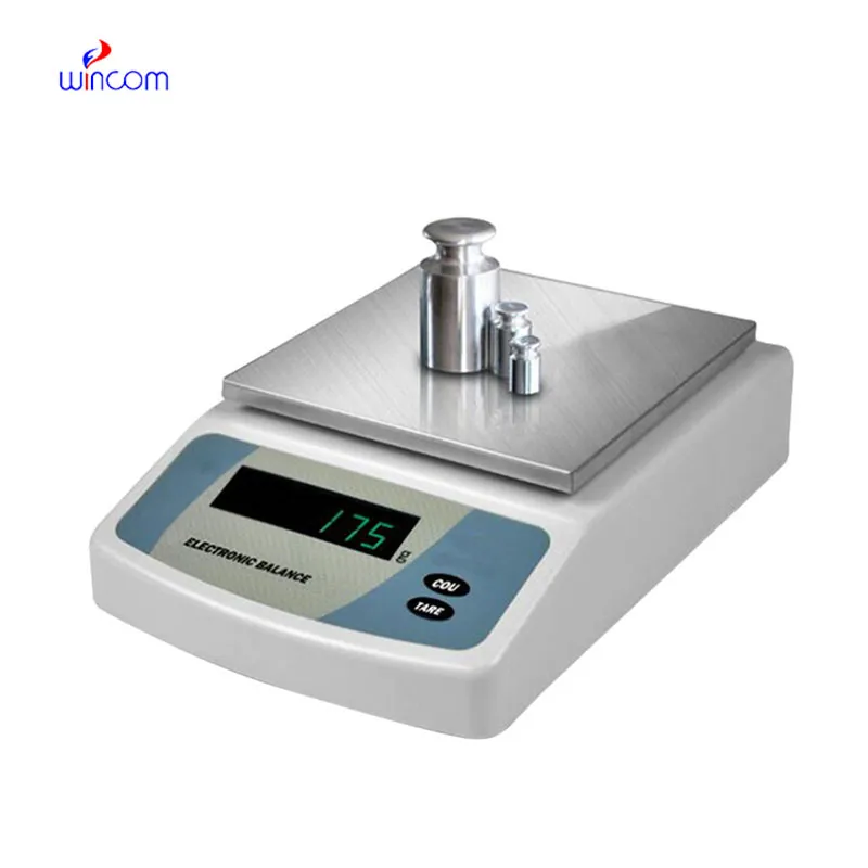
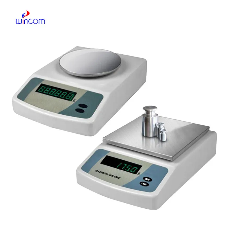
Q: What is the main purpose of an Analytical Balance? A: Its purpose is mainly to measure very tiny sample masses with the utmost precision in laboratories and hospitals. Q: What is the typical weighing range of an Analytical Balance? A: The weighing range for the majority of analytical balances is from 0 up to some grams with a resolution of micrograms or milligrams. Q: What environmental controls are necessary for an Analytical Balance's operation? A: Airflow, vibration, and temperature changes should not only be avoided but also prevented in the room where the scale is situated. Q: Is an Analytical Balance permitted in a hospital laboratory? A: Yes, it has indeed found widespread usage for the preparation of reagents, calibra¬tion, and drug development applications. Q: What should be the frequency of calibration for an Analytical Balance? A: The calibration interval is subject to the degree of use and the particular laboratory requirements.
We’ve used this centrifuge for several months now, and it has performed consistently well. The speed control and balance are excellent.
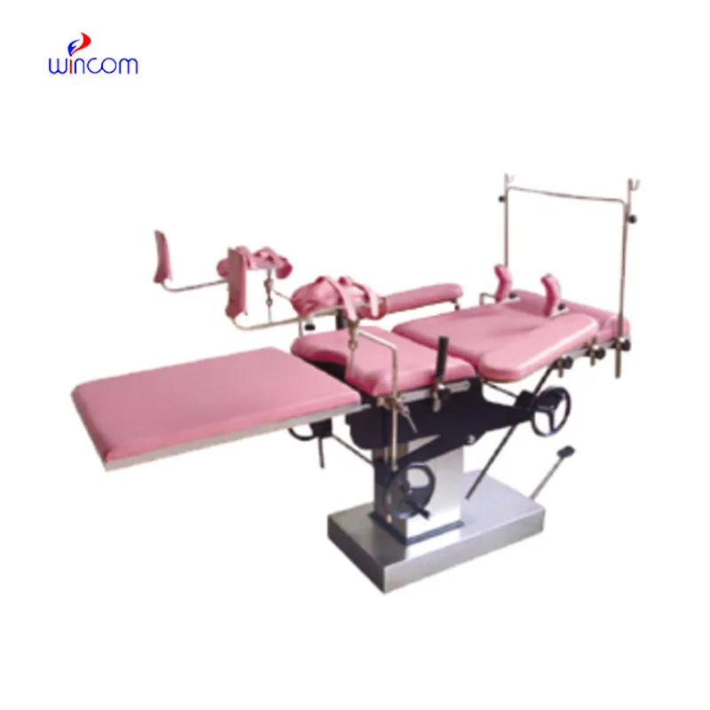
The delivery bed is well-designed and reliable. Our staff finds it simple to operate, and patients feel comfortable using it.
To protect the privacy of our buyers, only public service email domains like Gmail, Yahoo, and MSN will be displayed. Additionally, only a limited portion of the inquiry content will be shown.
I’m looking to purchase several microscopes for a research lab. Please let me know the price list ...
Hello, I’m interested in your water bath for laboratory applications. Can you confirm the temperat...
E-mail: [email protected]
Tel: +86-731-84176622
+86-731-84136655
Address: Rm.1507,Xinsancheng Plaza. No.58, Renmin Road(E),Changsha,Hunan,China

 af
af
 es
es
 ar
ar
 tr
tr
 sw
sw
 pt
pt
 th
th
 ur
ur
 bn
bn
 ne
ne
 vi
vi
 km
km
 lo
lo
 de
de
 ru
ru
 fi
fi
 nl
nl
 fa
fa
 fr
fr
 ko
ko