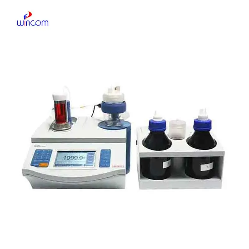
The use of electronic balancing in pharmaceutical laboratories is crucial for the accurate measurement of both active substances and excipients. Its extraordinary accuracy eliminates the possibility of formulation errors and makes regulatory compliance easier. electronic balancing are employed by laboratory staff for daily quality control, validation of batches, and research activities. Adding electronic balancing to the laboratory operations not only the consistency but also the reproducibility and the accuracy of the results for clinical trials and research applications are assured.

electronic balancing is commonly used in the compounding of medicinal and chemical substances in small quantities in hospital pharmacy laboratories. Mass control with high precision is critical when dealing with active pharmaceutical ingredients in micro or milligram quantities. This application helps to prepare exact doses, internal validation, and experimental drug research. By allowing for repeatable measurements, electronic balancing helps pharmacists and researchers in controlling formulation processes, which makes it easier to get consistency and reliability throughout hospital medication development workflows.
The future usage of electronic balancing in hospitals will mainly be geared towards the compatibility of automation. It is predicted that analytical balances will connect with the robotic sample handling mechanisms of clinical and pharmaceutical labs. This connection will allow for uninterrupted work, lesser operator involvement, and better consistency. The scale of La due to the hospitals aiming for total automation, electronic balancing will be your basic measuring unit in these cutting-edge platforms.
For electronic balancing to last long, professionals must do scheduled servicing as per the laboratory guidelines. Internal cleaning and checking of parts will help to slow down and eventually stop the performance degradation. Hospitals or labs that maintain structured service intervals not only enjoy lesser downtimes but also higher accuracy, thus they can carry on with seamless analytical operations.
electronic balancing is of great importance for developing and validating laboratory methods. One of the most important prerequisites for getting correct analytical results is the weighing of reference standards, buffers, and chemicals with absolute accuracy. Laboratory personnel depend on electronic balancing for constant concentration reproduction and hence, reliable testing. Its superb sensitivity plus reliable performance indeed make it a primary instrument for validating methods in the clinical, hospital, and pharmaceutical lab settings.
Q: What is the impact of temperature on the performance of analytical balance? A: The changes in temperature can lead to drift and weighing inconsistency. Q: Are analytical balances the only ones used in research laboratories? A: They are very important also for other processes such as sample preparation and improving the accuracy of the experiment. Q: How long does it usually take for an analytical balance to warm up? A: Warm-up times differ from one model to another, but an adequate stabilizing period increases the reliability of the measurement. Q: Is it possible for analytical balances to save weighing data? A: Internal memory or external data transfer are the two ways in which many models can achieve this feature. Q: Would it be necessary to undergo training if one wants to operate an analytical balance? A: Basic laboratory training will be enough to make sure that the balance is being used correctly.
The centrifuge operates quietly and efficiently. It’s compact but surprisingly powerful, making it perfect for daily lab use.
The hospital bed is well-designed and very practical. Patients find it comfortable, and nurses appreciate how simple it is to operate.
To protect the privacy of our buyers, only public service email domains like Gmail, Yahoo, and MSN will be displayed. Additionally, only a limited portion of the inquiry content will be shown.
I’d like to inquire about your x-ray machine models. Could you provide the technical datasheet, wa...
We’re interested in your delivery bed for our maternity department. Please send detailed specifica...
E-mail: [email protected]
Tel: +86-731-84176622
+86-731-84136655
Address: Rm.1507,Xinsancheng Plaza. No.58, Renmin Road(E),Changsha,Hunan,China

 af
af
 es
es
 ar
ar
 tr
tr
 sw
sw
 pt
pt
 th
th
 ur
ur
 bn
bn
 ne
ne
 vi
vi
 km
km
 lo
lo
 de
de
 ru
ru
 fi
fi
 nl
nl
 fa
fa
 fr
fr
 ko
ko