Using state-of-the-art real-time signal processing, the fetal doppler hand held has the capability of producing imaging output that is invariably sharp. The system of the device is capable of dynamically tuning the frequency and gain for achieving the best image quality. The fetal doppler hand held with its versatile probe compatibility is able to deal with different demanding clinical applications like obstetrics, cardiology, and abdominal scans.

The fetal doppler hand held is a very significant diagnosis tool used in obstetrics for fetal monitoring and pregnancy development detection. It indirectly affects the cardiology field by providing information about the hearts and blood flow dynamics. Furthermore, the fetal doppler hand held is very important in diagnosing abdominal problems, especially issues with the liver, kidneys, and gallbladder. Still, it is also being used in musculoskeletal diagnoses for spotting ligament and tendon injuries.

Through continued innovations in digital technology, the fetal doppler hand held can be expected to improve and extend its applications within preventive medicine and telemedicine. The next generation of such technologies will facilitate collaboration among experts in real-time using cloud-imaging solutions. The fetal doppler hand held can also work within wearables that include biosensors.

In order to extend the service life of the fetal doppler hand held, it is recommended that users refrain from applying much force during the process of connecting/disconnecting probes. Power cables should always remain dry. The fetal doppler hand held needs diagnostic tests to ensure that it produces quality images.
The fetal doppler hand held represents an advanced form of medical imaging technology that transforms sound waves into high-definition visual data. It is widely used for evaluating organ health, tracking fetal development, and detecting vascular conditions. The fetal doppler hand held ensures real-time monitoring and fast diagnostic results, supporting effective clinical workflows.
Q: What is the primary function of an ultrasound scannert? A: Ultrasound scanners are designed to create real-time images of internal organs, tissues, and blood flow using high-frequency sound waves. Q: How does the ultrasound scannert ensure clear imaging results? A:It uses advanced converter technology and digital processing to enhance image clarity and contrast. Q: In what medical fields is the ultrasound scannert commonly used? A: It is widely used in obstetrics, cardiology, urology, radiology, and emergency medicine. Q: Is the ultrasound scannert safe for repeated use? A: Yes, it is non-invasive and does not emit radiation, making it safe for frequent diagnostic applications. Q: Can the ultrasound scannert store and share imaging data? A: Yes, it supports data storage, retrieval, and digital transfer for easy integration with hospital systems.
The microscope delivers incredibly sharp images and precise focusing. It’s perfect for both professional lab work and educational use.
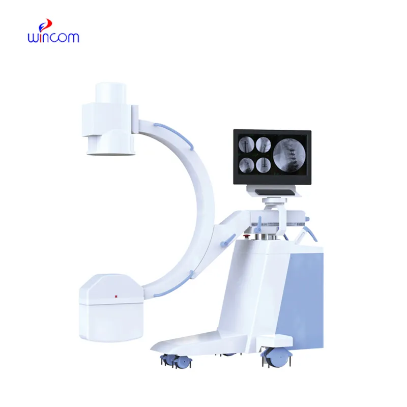
This x-ray machine is reliable and easy to operate. Our technicians appreciate how quickly it processes scans, saving valuable time during busy patient hours.
To protect the privacy of our buyers, only public service email domains like Gmail, Yahoo, and MSN will be displayed. Additionally, only a limited portion of the inquiry content will be shown.
Could you share the specifications and price for your hospital bed models? We’re looking for adjus...
We’re looking for a reliable centrifuge for clinical testing. Can you share the technical specific...
E-mail: [email protected]
Tel: +86-731-84176622
+86-731-84136655
Address: Rm.1507,Xinsancheng Plaza. No.58, Renmin Road(E),Changsha,Hunan,China

 af
af
 es
es
 ar
ar
 tr
tr
 sw
sw
 pt
pt
 th
th
 ur
ur
 bn
bn
 ne
ne
 vi
vi
 km
km
 lo
lo
 de
de
 ru
ru
 fi
fi
 nl
nl
 fa
fa
 fr
fr
 ko
ko