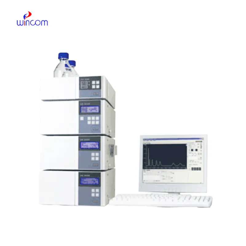
Today, clinical laboratories always rely on hplc column for the purpose of giving comprehensive chemical and biological data from patient samples. The technology's exceptional sensitivity and accuracy make it possible to separate even the smallest amounts of substances such as drugs and metabolites from complicated mixtures. Laboratory staff performs using hplc column in method development, validation and ongoing monitoring of the lab's analytical performance. The multi-use of the instrument guarantees its presence during both normal testing and research work, hence hospitals and laboratories are always consistent in providing accurate and trustworthy diagnostic and analytical results.

The quality control process for hplc column in intravenous medications and hospital-prepared solutions is being carried out by hospital laboratories. It isolates the impurities and analyzes the active substances to ascertain the uniformity of the composition. This practice enables the pharmacists and laboratory staff to verify the drug's quality before it gets to the patient, hence minimizing the risk associated with it and at the same time endorsing the safe therapeutic practices in hospitals.
In hospitals and clinical research, hplc column techniques will get higher resolution columns and ultrafast chromatography methods more and more. It will be possible to do these innovations in a shorter time and with a more accurate result. Future hplc column applications will be used to identify biomarkers quickly, monitor therapies in real-time, and manage patients more efficiently in both the laboratory and clinical settings.
Systematic attention on the system components is necessary for the running of hplc column in hospital and research labs. To prevent contamination and pressure problems, flushing of columns, seal replacements, and tubing inspections should be done regularly. Regular calibration of detectors and documentation of maintenance procedures should be done by laboratory technicians. The instruments' life is prolonged by consistent care and monitoring, which also lead to accurate sample analysis and support the reliability of laboratory operations both for clinical and experimental purposes.
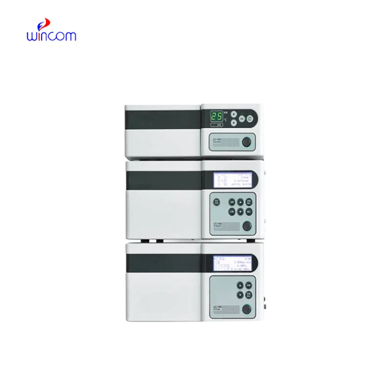
hplc column is of utmost importance in biochemistry laboratories of both universities and hospitals. It makes detailed study of proteins, peptides, and metabolites possible through the separation of intricate mixtures. The application of it includes but is not limited to enzymatic analysis, biomarker detection, and data obtained through metabolomics. The sensitivity and reproducibility of the device guarantee genuine molecular profiles. Lab technicians make use of hplc column to conclude their experiments and provide evidence for scientific publications. Its accuracy and versatility give biochemistry labs the ability to perform cutting-edge research in molecular mechanisms, disease pathways, and therapy targets thus, it becomes an indispensable tool for both analytical and clinical lab investigations.
Q: What is the sample preparation for HPLC? A: For the most part, samples should be filtered, diluted, or subjected to solvent extraction in order to avoid column clogs and have the results be accurate Q: Is HPLC able to pick trace-level compounds? A: With the right detectors, it can pick up such substances in extremely small amounts with high sensitivity. Q: Is HPLC a method that can be applied to analysis of proteins? A: Yes, particularly if one employs size-exclusion and reversed-phase columns for protein, peptide, and biomolecule separation. Q: What is the process of calibrating HPLC? A: The process is done by taking standards of known concentrations that are the same as the one in the sample and using them to check the performance of the column and the accuracy of the detector. Q: Are particular solvents needed for HPLC? A: Yes, the solvents used need to be compatible with the type of the column and the detectors to prevent any damage or interference in the analysis process.
The hospital bed is well-designed and very practical. Patients find it comfortable, and nurses appreciate how simple it is to operate.
I’ve used several microscopes before, but this one stands out for its sturdy design and smooth magnification control.
To protect the privacy of our buyers, only public service email domains like Gmail, Yahoo, and MSN will be displayed. Additionally, only a limited portion of the inquiry content will be shown.
Hello, I’m interested in your centrifuge models for laboratory use. Could you please send me more ...
Could you please provide more information about your microscope range? I’d like to know the magnif...
E-mail: [email protected]
Tel: +86-731-84176622
+86-731-84136655
Address: Rm.1507,Xinsancheng Plaza. No.58, Renmin Road(E),Changsha,Hunan,China

 af
af
 es
es
 ar
ar
 tr
tr
 sw
sw
 pt
pt
 th
th
 ur
ur
 bn
bn
 ne
ne
 vi
vi
 km
km
 lo
lo
 de
de
 ru
ru
 fi
fi
 nl
nl
 fa
fa
 fr
fr
 ko
ko