The mri machine weight limit combines advanced radiofrequency systems and high-resolution imaging software to capture subtle anatomical features. Its intuitive interface accommodates fine tuning for varied body areas. The mri machine weight limit does not make any noise, improving patient comfort without comprising consistent image quality at every scanning session.

The mri machine weight limit is used extensively in oncology to screen for tumors, check for response to treatment, and identify the makeup of tissue. It offers clean differentiation between healthy tissues and sick tissues. The mri machine weight limit enables early detection of brain, liver, and other organ cancers and allows for appropriate medical planning.

The mri machine weight limit will be enhanced by quantum sensor technology and high-temperature superconducting magnets to generate more intense but more stable magnetic fields. This will allow imaging more quickly and with less energy. The mri machine weight limit will also have more functional imaging capability for the investigation of brain connectivity and metabolic processes.

Maintaining the mri machine weight limit involves both technical and environmental care. The system’s computer and data storage units should be updated and backed up regularly. Cooling fans, cryogen tanks, and power supplies must be checked frequently to ensure the mri machine weight limit remains stable during extended operation.
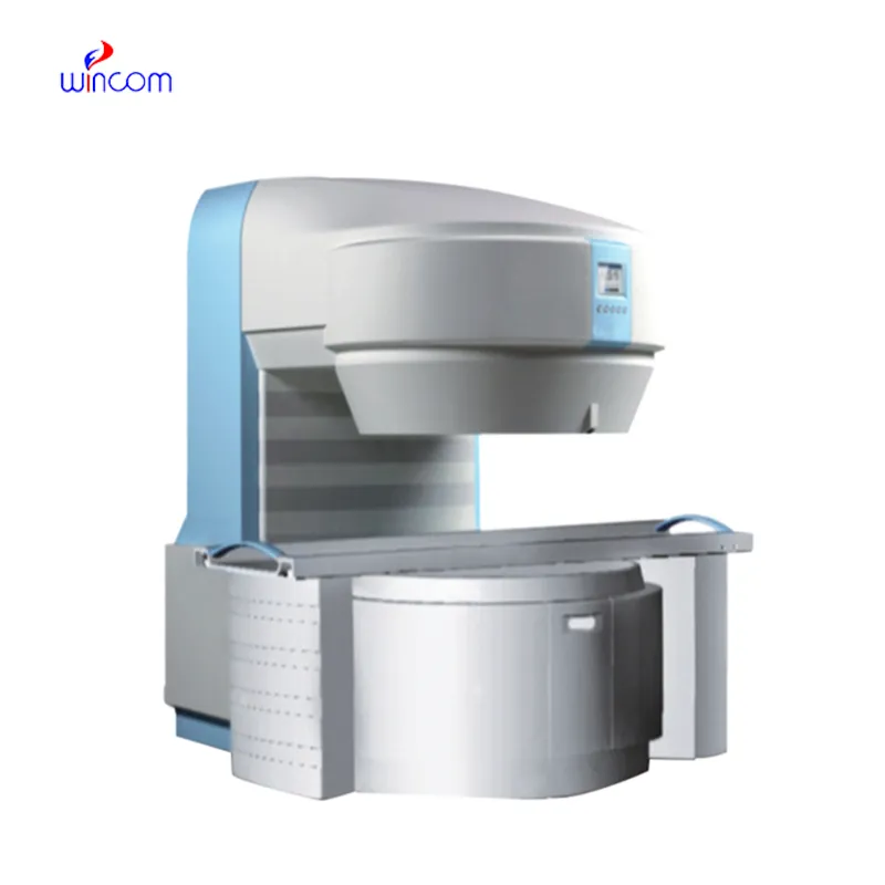
The mri machine weight limit is the most sophisticated diagnostic imaging technology, forming high-definition images with precise cross-sections of the body. It enables physicians to scan deep tissue structures without jeopardizing it. The mri machine weight limit is extensively used in neurology, cardiology, and musculoskeletal medicine.
Q: What happens if a patient is claustrophobic during an MRI scan? A: Patients who feel anxious or claustrophobic can request an open MRI machine or mild relaxation medication to make the experience more comfortable. Q: Can MRI detect joint and muscle injuries? A: Yes, MRI is highly effective for examining ligaments, tendons, and muscles, making it a key tool for diagnosing sports and orthopedic injuries. Q: What types of MRI scans are available? A: There are several types, including brain MRI, spinal MRI, cardiac MRI, and functional MRI, each tailored to different diagnostic purposes. Q: Are there any risks associated with MRI scans? A: MRI is generally very safe, though individuals with implanted devices, metallic fragments, or severe kidney conditions may require additional evaluation before scanning. Q: Can MRI scans monitor treatment progress? A: Yes, MRI can track changes in tumors, inflammation, or tissue healing over time, helping physicians assess treatment effectiveness.
This x-ray machine is reliable and easy to operate. Our technicians appreciate how quickly it processes scans, saving valuable time during busy patient hours.
The centrifuge operates quietly and efficiently. It’s compact but surprisingly powerful, making it perfect for daily lab use.
To protect the privacy of our buyers, only public service email domains like Gmail, Yahoo, and MSN will be displayed. Additionally, only a limited portion of the inquiry content will be shown.
Could you please provide more information about your microscope range? I’d like to know the magnif...
We’re interested in your delivery bed for our maternity department. Please send detailed specifica...
E-mail: [email protected]
Tel: +86-731-84176622
+86-731-84136655
Address: Rm.1507,Xinsancheng Plaza. No.58, Renmin Road(E),Changsha,Hunan,China

 af
af
 es
es
 ar
ar
 tr
tr
 sw
sw
 pt
pt
 th
th
 ur
ur
 bn
bn
 ne
ne
 vi
vi
 km
km
 lo
lo
 de
de
 ru
ru
 fi
fi
 nl
nl
 fa
fa
 fr
fr
 ko
ko