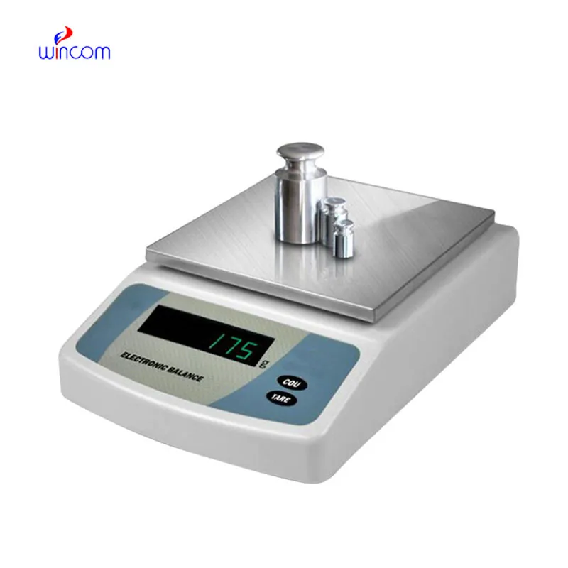
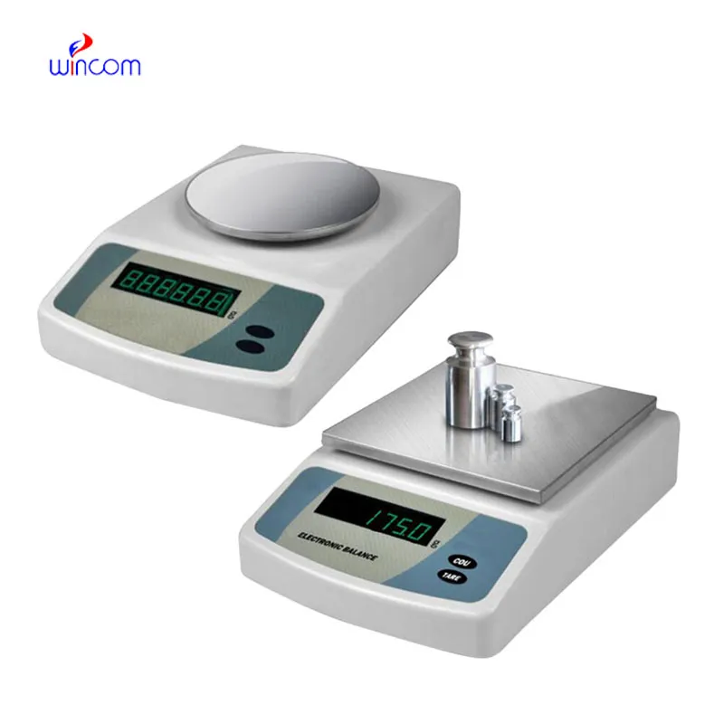
sartorius analytical balance is an extremely accurate device that is specifically designed for weighing little amounts in labs and hospital pharmacies. The instrument's sensitivity implies accurate preparation of samples for diagnostic testing, reagent making, and the production of drugs. The lab personnel consider it vital to get repeatable measurements, perform calibration checks, and validate their standards using sartorius analytical balance. The right use of sartorius analytical balance will bring about the smoothness of clinical workflows, research activities, and quality control, thus providing accuracy and reliability to all analytical processes in hospitals and laboratories.

The use of sartorius analytical balance comes in hospital toxicology laboratories, where they have to do analytical testing with trace-level substances. First, accurate weighing is necessary before the sample is processed and detected by the instrument. This application ensures accurate quantification and repeatable testing conditions, especially when working with very small sample volumes. By maintaining mass consistency, sartorius analytical balance contributes to the reliability of the toxicological analysis meant for clinical assessment and research.
sartorius analytical balance will envision a new era where the user-centered interfaces of hospital workers take greater priority. Such friendly visualizations, accompanied by the directing of workflows and notifications, will prove to be helpful during the working of professionals with machines. This change will cut down the duration of training and will also bring in better quality of work in clinical laboratories. sartorius analytical balance will not give up on the accuracy aspect while still allowing the ease of use in the hospital environments that are changing fast.
The maintenance of sartorius analytical balance involves the aspects of storage and inactivity care that come first. The balance should be protected from dust and vibration when it is not in active use. Periodically checking the operational status during long storage prevents unnoticed performance drift. These practices guarantee that sartorius analytical balance is still capable of accurate use in laboratories, medical and hospital settings.
sartorius analytical balance plays an important role in the hospital pharmacy in the accurate formulation of medications, intravenous solutions, and compounded drugs. Even slight alterations in the weight of drugs can change the effectiveness of the drug and endanger the patient's safety. The Pharmacy Technicians rely on sartorius analytical balance for the correct dosing and checking of the active ingredients. The tool's accuracy guarantees the dependable preparation of drugs, the observance of the rules, and the quality control of the overall hospital pharmacy activities.
Q: What industries are the widespread users of Analytical Balances? A: Their primary application lies in laboratories, hospitals, pharmaceutical facilities, and research institutions. Q: Is it possible to measure liquids with an Analytical Balance? A: Liquids can be indirectly measured using appropriate containers. Q: What does it mean by repeatability in an Analytical Balance? A: It is the capability to provide constant results when tested under the same conditions. Q: Is it necessary for the installation of Analytical Balances to be in controlled environments? A: Controlled environments are beneficial for keeping accuracy and stability long term. Q: What is the average lifespan of an Analytical Balance? A: If taken care of and maintained properly, it will be a reliable and many years-long operating device.
We’ve been using this mri machine for several months, and the image clarity is excellent. It’s reliable and easy for our team to operate.
This x-ray machine is reliable and easy to operate. Our technicians appreciate how quickly it processes scans, saving valuable time during busy patient hours.
To protect the privacy of our buyers, only public service email domains like Gmail, Yahoo, and MSN will be displayed. Additionally, only a limited portion of the inquiry content will be shown.
Could you please provide more information about your microscope range? I’d like to know the magnif...
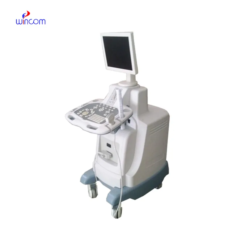
We’re currently sourcing an ultrasound scanner for hospital use. Please send product specification...
E-mail: [email protected]
Tel: +86-731-84176622
+86-731-84136655
Address: Rm.1507,Xinsancheng Plaza. No.58, Renmin Road(E),Changsha,Hunan,China

 af
af
 es
es
 ar
ar
 tr
tr
 sw
sw
 pt
pt
 th
th
 ur
ur
 bn
bn
 ne
ne
 vi
vi
 km
km
 lo
lo
 de
de
 ru
ru
 fi
fi
 nl
nl
 fa
fa
 fr
fr
 ko
ko