Built from high-quality optics, the sem microscope vs tem provides higher clarity for scientific and educational use. The durable body provides stable operation, and the adjustable head and stage setup provide ergonomic convenience. Advanced illumination systems enable observation with high contrast of transparent and reflected samples. The sem microscope vs tem is compatible with digital cameras and display devices, enabling real-time observation and recording of microscopic structures for further study and analysis.

The sem microscope vs tem has a wide range of professional and academic uses. In biomedical labs, it is used to analyze cell morphology and identify abnormalities. Industrial scientists rely on the sem microscope vs tem in testing product consistency, micro defect detection, and surface characterization. In agriculture, it is used to study plant diseases, seed morphology, and pest interactions. Museums and conservation centers apply the sem microscope vs tem in analyzing artwork materials to ensure proper preservation and restoration of historical works.

The sem microscope vs tem of the future will integrate optical engineering and computational imaging. Quantum sensors and nanophotonic devices will enable researchers to image at atomic levels. Smart automation will streamline workflow, where researchers read instead of physically setting. The sem microscope vs tem will use augmented reality interfaces, giving users direct access to multi-layered information. Through sustained innovation, it will be at the forefront of health science research, materials research, and environmental research.

A well-maintained sem microscope vs tem gives reliable performance and long operating life. Check optical elements regularly for dust, fingerprint, or oil residue. Use only authorized manufacturer cleaning materials to prevent lens coating damage. Store the sem microscope vs tem upright, supported, and covered when not in use. Check focusing mechanisms for smooth operation and illumination system for uniform brightness. Standard maintenance procedures minimize downtime and preserve imaging quality for education and research.
With a sem microscope vs tem, human man can explore the microcosm with unprecedented clarity. The instrument magnifies small samples so that exact study can be conducted in laboratories, clinics, and schools. The sem microscope vs tem recognizes cell morphology, bacterial cultures, and intricate material surfaces. Although optical and electronic technology has been enhanced, the sem microscope vs tem of today's time offers more magnification, image stability, and integration into digital media for efficient data registration and perception.
Q: What are the main parts of a microscope? A: The key components include the eyepiece, objective lenses, stage, focusing knobs, and illumination system, all working together to magnify and clarify specimens. Q: How do you clean the lenses of a microscope? A: Lenses should be cleaned using soft lens paper or microfiber cloth with a small amount of lens cleaner to avoid scratching or damaging optical coatings. Q: What magnification levels can a microscope achieve? A: Depending on the model, a microscope can typically achieve magnifications ranging from 40x to over 1000x for detailed observation of microscopic structures. Q: Why is light adjustment important in a microscope? A: Proper light adjustment ensures accurate contrast and brightness, allowing clear observation without distortion or glare during viewing. Q: Can a microscope be used for educational purposes? A: Yes, microscopes are widely used in classrooms and laboratories to teach students about biology, materials science, and microscopic analysis.
This x-ray machine is reliable and easy to operate. Our technicians appreciate how quickly it processes scans, saving valuable time during busy patient hours.
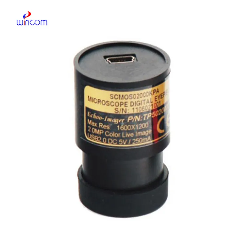
I’ve used several microscopes before, but this one stands out for its sturdy design and smooth magnification control.
To protect the privacy of our buyers, only public service email domains like Gmail, Yahoo, and MSN will be displayed. Additionally, only a limited portion of the inquiry content will be shown.
Hello, I’m interested in your centrifuge models for laboratory use. Could you please send me more ...
Could you please provide more information about your microscope range? I’d like to know the magnif...
E-mail: [email protected]
Tel: +86-731-84176622
+86-731-84136655
Address: Rm.1507,Xinsancheng Plaza. No.58, Renmin Road(E),Changsha,Hunan,China

 af
af
 es
es
 ar
ar
 tr
tr
 sw
sw
 pt
pt
 th
th
 ur
ur
 bn
bn
 ne
ne
 vi
vi
 km
km
 lo
lo
 de
de
 ru
ru
 fi
fi
 nl
nl
 fa
fa
 fr
fr
 ko
ko