The ultrasound ground scanner has been designed considering the needs of modern health care, providing uninterrupted performance with its rapid image acquisition and high-definition visualization. The robust outer casing and sophisticated temperature control system will make sure that the device continues to work and be trustworthy. Also, the ultrasound ground scanner is a device that aids the long-term data archiving process for efficient medical record management.

The ultrasound ground scanner is a tool that medical professionals are using in different departments like pediatrics for the measurement of the size of the organs and neurology for appreciating the anatomy of the soft tissues. It makes it easy for anesthesiologists to perform nerve blocks and vascular access procedures. The ultrasound ground scanner increase the speed and the trust of the diagnosis during both routine checkups and highly technical interventions.

In addition, as new technologies emerge, the ultrasound ground scanner is expected to become more compact and intelligent with better diagnostic capabilities. The new ultrasound ground scanner will incorporate 3D and 4D capabilities. The ultrasound ground scanner will also be integrated with digital hospitals for seamless management of patient data.

Care of the ultrasound ground scanner involves much more than cleanup. Environmental monitoring and mechanical protection are also part of the process. The ultrasound ground scanner should not be subject to either vibration or shock. The ultrasound ground scanner should be backed up periodically to retain vital images.
Built for performance and accuracy, the ultrasound ground scanner is an imaging diagnosis platform. It gives real-time images of tissues, organs, and vascular systems, enabling increased detection of pathology. Space-efficient and compact, the ultrasound ground scanner is ideal for hospitals, clinics, and ambulatory healthcare facilities. Its accuracy imaging enables physicians to offer timely and informed medical care.
Q: How does the ultrasound scannert contribute to emergency diagnostics? A: It enables rapid assessment of internal injuries and organ conditions in time-sensitive situations. Q: Can the ultrasound scannert be upgraded with new features? A: Yes, most models support software updates to enhance performance and expand diagnostic functions. Q: What kind of power supply does the ultrasound scannert use? A: It operates on standard AC power and may include rechargeable battery options for mobile use. Q: Is the ultrasound scannert compatible with electronic medical record systems? A: Yes, it can connect to EMR systems to streamline patient data entry and storage. Q: What factors influence the image quality of the ultrasound scannert? A: Image quality depends on probe type, operator technique, and the frequency settings selected for scanning.
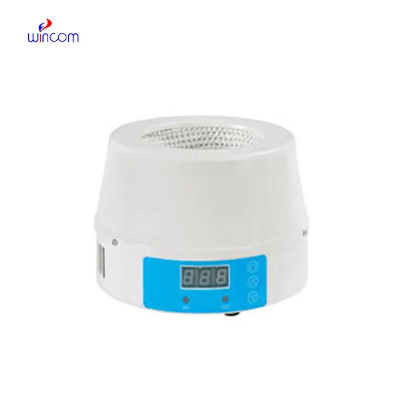
The water bath performs consistently and maintains a stable temperature even during long experiments. It’s reliable and easy to operate.
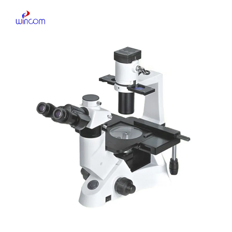
I’ve used several microscopes before, but this one stands out for its sturdy design and smooth magnification control.
To protect the privacy of our buyers, only public service email domains like Gmail, Yahoo, and MSN will be displayed. Additionally, only a limited portion of the inquiry content will be shown.
Could you share the specifications and price for your hospital bed models? We’re looking for adjus...
We’re currently sourcing an ultrasound scanner for hospital use. Please send product specification...
E-mail: [email protected]
Tel: +86-731-84176622
+86-731-84136655
Address: Rm.1507,Xinsancheng Plaza. No.58, Renmin Road(E),Changsha,Hunan,China

 af
af
 es
es
 ar
ar
 tr
tr
 sw
sw
 pt
pt
 th
th
 ur
ur
 bn
bn
 ne
ne
 vi
vi
 km
km
 lo
lo
 de
de
 ru
ru
 fi
fi
 nl
nl
 fa
fa
 fr
fr
 ko
ko