Designed with efficiency in mind, the veterinary x ray machines combines exposure control and image processing. The equipment enables the observation of detail in both the bone and soft tissues without much distortion. The veterinary x ray machines offers high flexibility when it comes to operations and can work well in various settings like hospitals and research labs.

In veterinary practice, the veterinary x ray machines analyzes bone fractures, joint diseases, and internal ailments in animals. It provides a transparent image of bone and organ structure, allowing veterinarians to observe health condition precisely. The veterinary x ray machines is extremely important in animal health care and research.

In the years to come, the veterinary x ray machines shall be at the forefront of predictive and personalized medicine. Smart imaging platforms shall look into patient history in conjunction with real-time scans in an attempt to predict upcoming health problems. The veterinary x ray machines shall therefore turn into an anticipatory diagnostic system instead of a reactive one.

Maintenance of the veterinary x ray machines requires close attention to mechanical, electrical, and imaging parts. Regular visual examination catches wear or damage early. The veterinary x ray machines must be cleaned using non-abrasive substances, and filters or protective covers periodically replaced. Preventive maintenance minimizes downtime and provides reliable diagnostic results.
The veterinary x ray machines turns X-ray radiation into visual information that offers clarity on skeletal and soft tissues. The digital imaging technology of the veterinary x ray machines helps improve the clarity of images by reducing radiation. The veterinary x ray machines helps healthcare providers evaluate a patient's skeletal system since it assists in diagnosing fractures, lung disease, and dental problems.
Q: What makes an x-ray machine different from a CT scanner? A: An x-ray machine captures a single 2D image, while a CT scanner takes multiple x-rays from different angles to create 3D cross-sectional views. Q: How is image quality measured in an x-ray machine? A: Image quality depends on factors like contrast, resolution, and exposure settings, which are adjusted based on the target area being examined. Q: What power supply does an x-ray machine require? A: Most x-ray machines operate on high-voltage power systems, typically between 40 to 150 kilovolts, depending on their intended use. Q: Can x-ray machines be used for dental imaging? A: Yes, specialized dental x-ray machines provide detailed images of teeth, jaws, and surrounding structures to support oral health assessments. Q: How does digital imaging improve x-ray efficiency? A: Digital systems allow instant image preview, faster diagnosis, and reduced need for retakes, improving workflow efficiency in clinical environments.
The microscope delivers incredibly sharp images and precise focusing. It’s perfect for both professional lab work and educational use.
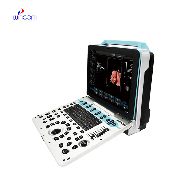
This ultrasound scanner has truly improved our workflow. The image resolution and portability make it a great addition to our clinic.
To protect the privacy of our buyers, only public service email domains like Gmail, Yahoo, and MSN will be displayed. Additionally, only a limited portion of the inquiry content will be shown.
Hello, I’m interested in your water bath for laboratory applications. Can you confirm the temperat...
Hello, I’m interested in your centrifuge models for laboratory use. Could you please send me more ...
E-mail: [email protected]
Tel: +86-731-84176622
+86-731-84136655
Address: Rm.1507,Xinsancheng Plaza. No.58, Renmin Road(E),Changsha,Hunan,China

 af
af
 es
es
 ar
ar
 tr
tr
 sw
sw
 pt
pt
 th
th
 ur
ur
 bn
bn
 ne
ne
 vi
vi
 km
km
 lo
lo
 de
de
 ru
ru
 fi
fi
 nl
nl
 fa
fa
 fr
fr
 ko
ko