Compact but powerful, the virus under microscope delivers advanced control for detailed visual analysis. The precision-designed optical pathway and coated lenses reduce glare and optimize image quality. The adjustable stage accommodates multiple slide sizes, and the light source offers steady brightness for long observation periods. The virus under microscope is built for durability with low maintenance and reliable operation in high laboratory throughput. It is ideal for professional and academic applications requiring accurate visual results.

The virus under microscope is applied widely in biology for studying cells, tissues, and microorganisms with unmatched clarity. Clinically, it is applied to assist in the diagnosis of infections, blood diseases, and cell disorders. In industry, the virus under microscope is employed for material examination, surface flaw detection, and microstructure analysis of metals and polymers. In institutions of learning, it is a teaching tool that helps students learn microscopic anatomy and chemical reactions. Its use extends into environmental monitoring where it is used to analyze soil or water samples to ascertain quality and detect pollutants.

In the short term, the virus under microscope will be a networked and completely digital platform. Integration with AI-powered recognition systems will make automated cell, material, and organism recognition possible. Cloud storage will allow easier information sharing and archiving. The virus under microscope is set to embrace holographic and super-resolution techniques, allowing researchers to see structures at the molecular scale. This technology will open new fields in diagnostics, nanoscience, and education, which will expand the use of microscopic observation across industries.

Preventive maintenance ensures the virus under microscope operate reliably for years. Clean all glass surfaces gently to avoid abrasion. Moving parts, including the stage and focusing devices, need to be cleaned for dust and adjusted to run smoothly. The virus under microscope need to be placed on a vibration-free surface so that internal alignment is not compromised. Power cords and switches also need to be checked so that no electrical damage is caused. Periodic servicing by an expert keeps the optical components centered and in balance precisely.
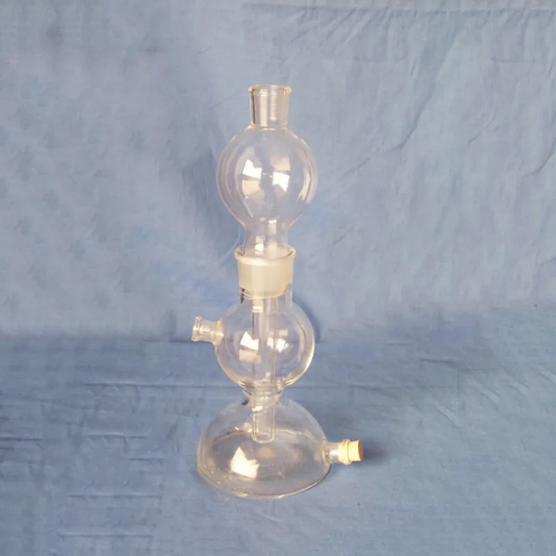
A virus under microscope is an essential investigation device that brings new insight to life in material and biological science. It magnifies objects which are too small to view with the naked eye, revealing hidden patterns and structures of cells. The virus under microscope facilitates proper observation by possessing fine optics, variable illumination, and fine focusing devices. It plays an important part in laboratory testing, forensic analysis, and industrial quality control, revealing vivid insights into the structure and behavior of microscopic material.
Q: What is a microscope used for? A: A microscope is used to magnify tiny objects or structures, allowing detailed observation of cells, microorganisms, and materials that are invisible to the naked eye. Q: How often should a microscope be calibrated? A: To maintain measurement accuracy and ensure accurate focus during research or analysis, regular calibration should be performed, typically once or twice a year. Q: What type of light source is commonly used in a microscope? A: Most modern microscopes use LED or halogen light sources, which provide stable light and adjustable brightness for clear images at a wide range of magnifications. Q: Can a microscope be connected to a computer? A: Yes, many microscope models feature USB or HDMI ports that allow image capture and digital display through specialized imaging software. Q: How should a microscope be stored when not in use? A: A microscope should be covered with a dust shield and stored in a cool, dry location to prevent contamination and protect optical components from humidity.
This x-ray machine is reliable and easy to operate. Our technicians appreciate how quickly it processes scans, saving valuable time during busy patient hours.
This ultrasound scanner has truly improved our workflow. The image resolution and portability make it a great addition to our clinic.
To protect the privacy of our buyers, only public service email domains like Gmail, Yahoo, and MSN will be displayed. Additionally, only a limited portion of the inquiry content will be shown.
Hello, I’m interested in your centrifuge models for laboratory use. Could you please send me more ...
We’re looking for a reliable centrifuge for clinical testing. Can you share the technical specific...
E-mail: [email protected]
Tel: +86-731-84176622
+86-731-84136655
Address: Rm.1507,Xinsancheng Plaza. No.58, Renmin Road(E),Changsha,Hunan,China

 af
af
 es
es
 ar
ar
 tr
tr
 sw
sw
 pt
pt
 th
th
 ur
ur
 bn
bn
 ne
ne
 vi
vi
 km
km
 lo
lo
 de
de
 ru
ru
 fi
fi
 nl
nl
 fa
fa
 fr
fr
 ko
ko