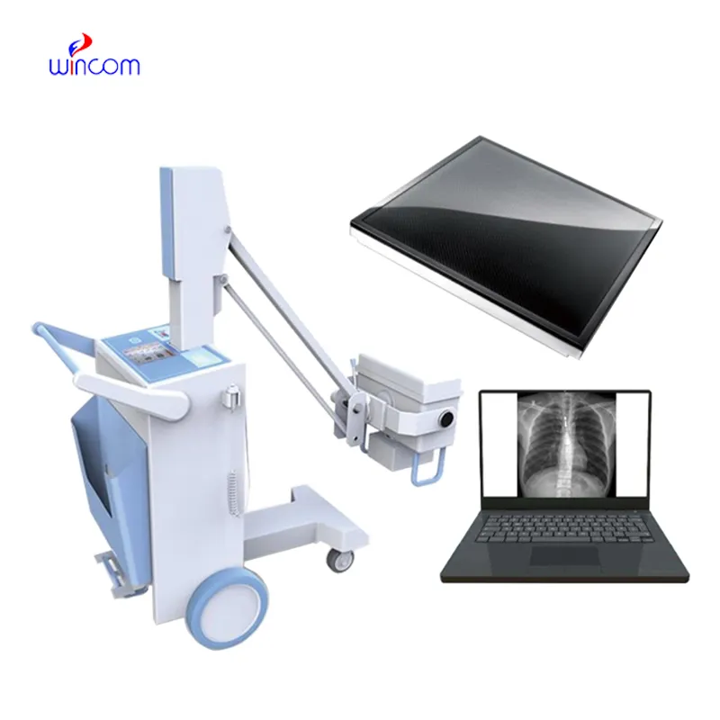
The x ray baggage scanner comes equipped with advanced digital detectors that transform X-ray energy into high definition images of incredible detail. The system's design makes it easy to use and facilitates quick image capturing. The x ray baggage scanner system can be connected effortlessly to hospital information systems that enable the secure transfer of data. The system's robust design provides support for long-term use within healthcare settings.

The x ray baggage scanner is used in numerous various clinical departments, including orthopedics, radiology, and oncology. It is usually applied to detect lung lesions and bone fractures as well as track tumor growth. In children's practice, the x ray baggage scanner enables safe imaging with child-friendly doses suitable for diagnostic needs in children.

The next generation of the x ray baggage scanner would be oriented toward digital transformation through intelligent image algorithms. Machine learning would enhance the pace of image reconstruction and image clarity. The x ray baggage scanner would be made wireless and portable to enable remote diagnosis and mobile healthcare facilities.

Regular upkeep of the x ray baggage scanner enhances operating efficiency and patient safety. Regular exposure level checks and image acuteness tests guarantee reproducible output. The x ray baggage scanner should be operated by well-trained operators who observe cleaning and handling protocols to reduce wear and prolong service life.
The x ray baggage scanner works by providing X-rays that penetrate the body to generate contrast images. The device has been extensively used for screening, diagnosing, and treatment. The x ray baggage scanner increases efficiency in healthcare by offering quick images for immediate analysis by a medical practitioner.
Q: What makes an x-ray machine different from a CT scanner? A: An x-ray machine captures a single 2D image, while a CT scanner takes multiple x-rays from different angles to create 3D cross-sectional views. Q: How is image quality measured in an x-ray machine? A: Image quality depends on factors like contrast, resolution, and exposure settings, which are adjusted based on the target area being examined. Q: What power supply does an x-ray machine require? A: Most x-ray machines operate on high-voltage power systems, typically between 40 to 150 kilovolts, depending on their intended use. Q: Can x-ray machines be used for dental imaging? A: Yes, specialized dental x-ray machines provide detailed images of teeth, jaws, and surrounding structures to support oral health assessments. Q: How does digital imaging improve x-ray efficiency? A: Digital systems allow instant image preview, faster diagnosis, and reduced need for retakes, improving workflow efficiency in clinical environments.
The centrifuge operates quietly and efficiently. It’s compact but surprisingly powerful, making it perfect for daily lab use.
We’ve been using this mri machine for several months, and the image clarity is excellent. It’s reliable and easy for our team to operate.
To protect the privacy of our buyers, only public service email domains like Gmail, Yahoo, and MSN will be displayed. Additionally, only a limited portion of the inquiry content will be shown.
I’d like to inquire about your x-ray machine models. Could you provide the technical datasheet, wa...
Hello, I’m interested in your centrifuge models for laboratory use. Could you please send me more ...
E-mail: [email protected]
Tel: +86-731-84176622
+86-731-84136655
Address: Rm.1507,Xinsancheng Plaza. No.58, Renmin Road(E),Changsha,Hunan,China

 af
af
 es
es
 ar
ar
 tr
tr
 sw
sw
 pt
pt
 th
th
 ur
ur
 bn
bn
 ne
ne
 vi
vi
 km
km
 lo
lo
 de
de
 ru
ru
 fi
fi
 nl
nl
 fa
fa
 fr
fr
 ko
ko